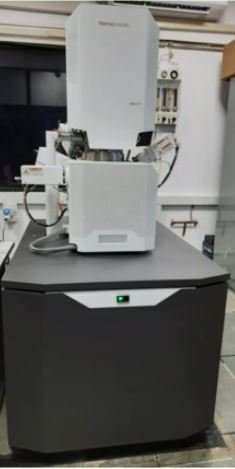
Field Emission Scanning Electron Microscopy (FESEM)
Field Emission Scanning Electron Microscopy (FESEM)
In a FESEM, electrons are liberated from a field emission source. The generated primary electrons accelerate in a high electrical field gradient. The primary electrons are focussed and deflected by electronic lenses to produce a narrow scan beam that bombards the object within the high vacuum column. The detector catches the secondary electrons containing the sample information and produces an electronic signal. This signal is amplified and transformed to a digital image that can be used for further studies by the user.
The Apreo 2 S SEM has the Thermo Scientific SmartAlign Technology, an optics system that aligns itself. Furthermore, the Apreo 2 S SEM automates the image fine-tuning process with Thermo Scientific FLASH technology which does necessary corrections to lens centering, the stigmators, and final focus of the image. The instrument has the below specifications.
FEG filament with better than 1.2 nm resolution.
Accelerating Voltage Range: 200 V – 30 kV.
EDS detector (Thermofisher, 30 mm2 with 127 eV spectral resolution)
In-lens, Secondary electron and low vacuum detectors.