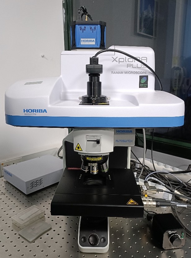
Raman spectroscopy is an analytical technique where scattered light is used to measure the vibrational energy modes of a sample. It is named after the Indian physicist C. V. Raman who, together with his research partner K. S. Krishnan, was the first to observe Raman scattering in 1928. Raman spectroscopy can provide both chemical and structural information, as well as the identification of substances through their characteristic Raman ‘fingerprint’. Raman spectroscopy extracts this information through the detection of Raman scattering from the sample.
Make: Horiba Scientific
Model: Xplora plus V1.2 Multiline
Research grade optical microscope : Olympus BX, complete microscope with 2 position motorized white light illuminator Koehler illumination by reflection (LED eqv 100 W)/ transmission (Halogen 30 W), Abbe condenser and 2 objectives
Lens: 10X, 20X, 50X, 100X
Integrated imaging spectrometer with 4 gratings mounted on motorized turret with gratings: 600gr, 1200gr, 1800gr and 2400gr HORIBA Scientific CCD detector, TE air-cooled (-60 ⁰ C), 1024×256 pixel, 16 bit
Motorized PC controlled 6 position ND filter wheel for laser power adjustment (0.1%, 1%, 10%, 25%, 50%, 100%)
LabSpec6 spectral software suite for the easy acquisition and analysis and analysis of Raman data. Includes control of the hardware and acquisition parameters, AUTO calibration, customizable methods, FLAT fluorescence subtraction, peak label and fit, image capture, smoothing, spectral subtraction etc.
Capabilities: Acquisition of point Raman spectra through confocal microscope from 50-3000 cm-1 at three excitation wavelengths: 532, 638 and 785 nm; Real time display of spectra; Powder, thin film and liquid samples; Mapping of Raman spectral lines across features